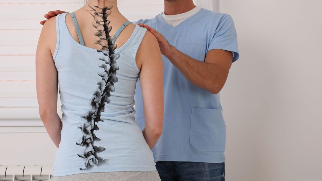
Elbow dislocation is most often a posterolateral displacement of the ulna and radius from the humerus, usually after a fall on an outstretched hand. Simple dislocations (no major fracture) are usually treated with prompt reduction, short‑term immobilisation, and early mobilisation, while complex dislocations with fractures often need surgery and longer rehabilitation.
Elbow dislocation is common in both children and adults. In children, it is termed as pulled elbow in children. It usually occurs when we fall on our outstretched hand.
In this article, we will cover elbow dislocation in detail. Will we learn the elbow dislocation anatomy, its classifications? How can we diagnose it? What is its treatment?
Anatomy of elbow joint dislocation
The elbow joint is formed by the articular surface of the humerus and upper articular surface of the ulna and the radius. So these three together form the elbow joint, and it gets dislocated.
An elbow dislocation can be simple, involving only a dislocation of the elbow, or complex, involving associated fractures.
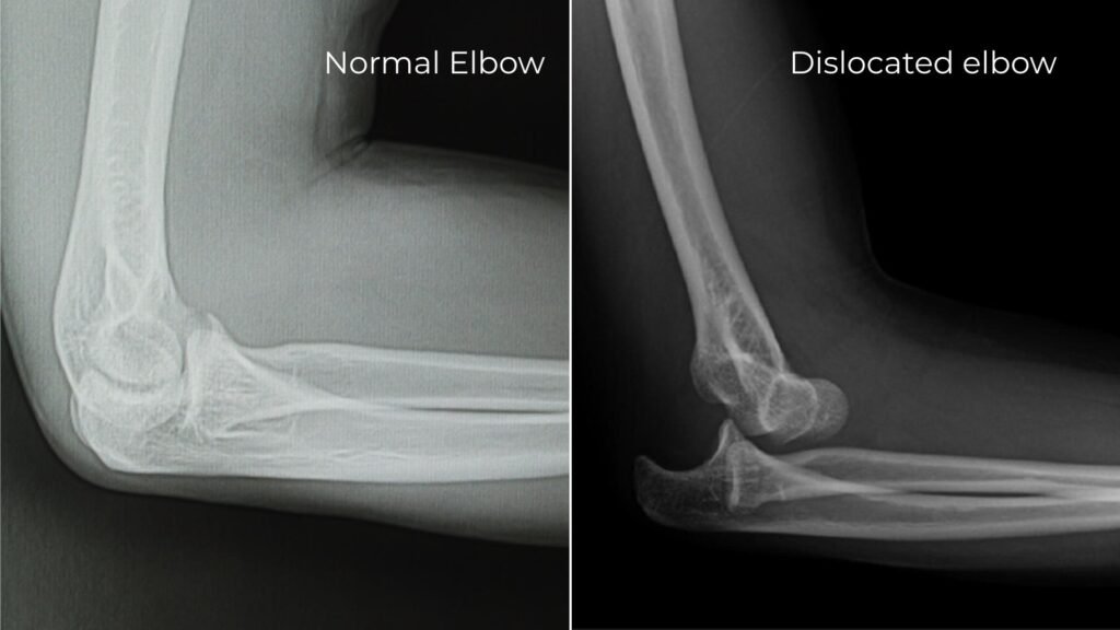
If you observe the X-ray, the one on the left is of a normal elbow joint, the one on the right is the elbow joint, where the elbow is dislocated, and it’s displaced posteriorly.
Simple vs complex elbow dislocation
| Feature | Simple elbow dislocation | Complex elbow dislocation |
|---|---|---|
| Definition | Elbow joint is dislocated without major associated fractures; injury mainly involves capsuloligamentous and soft‑tissue structures2. | Elbow joint is dislocated with one or more significant periarticular fractures, commonly of the radial head, coronoid process, or olecranon. |
| Associated fractures | No clinically important fractures around the elbow, or only minor avulsion fragments without mechanical impact on stability. | Fractures of radial head/neck, coronoid process, olecranon, distal humerus or combination injury patterns (e.g., “terrible triad”). |
| Typical treatment | Closed reduction, short‑term immobilisation (usually ≤2–3 weeks) followed by early active range‑of‑motion and progressive rehabilitation. | Often requires operative fixation or ligament reconstruction plus post‑operative immobilisation, then carefully protected rehabilitation to restore motion and stability2. |
| Prognosis | Generally excellent with early mobilisation: good pain relief, near‑normal range of motion, and low risk of recurrent instability in most patients2. | Prognosis more guarded: higher risk of stiffness, residual instability, post‑traumatic arthritis, and need for prolonged rehab, especially in complex fracture patterns. |
Types of elbow dislocation

So, a posterior dislocation is one type of elbow dislocation. There are other types as well, which leads us to the next title, the common types of dislocation1.
- Posterior dislocation: The most common is posterior dislocation.
- The second is posterior lateral, and
- The third is posterior medial.
Let me explain everything to you with a diagram. Look at the diagram with the humerus bone, the ulna, and the radius bone. In the first figure from the right, you can see that the ulna bone is dislocated, and it is displaced posteriorly.
That’s why we call this a posterior dislocation.
This was our lateral view. But if we look at this in an anterior-posterior view, the dislocation will look something like the one adjacent to the posterior dislocation.
Just left to it is the postero-medial dislocation. In this case, there is a posterior dislocation, and it is also displaced medially. That’s why we call it a posterior medial dislocation.
The third type is a posterior dislocation, and the dislocated segment is displaced laterally. So, we call this a posterior lateral dislocation.
Got it?
There are three types elbow dislocation: Posterior dislocation, postero-medial dislocation, and postero-lateral dislocation.
What is the mechanism of injury?
Like I told you in the introduction, a fall on the point of the elbow. When you fall and you land on the point of your elbow, there’s a chance of an elbow dislocation.
Or there could be a direct injury. A direct hit there can also cause elbow dislocation.
What are its clinical features?
There will be
- Pain.
- Swelling around the elbow.
- Tenderness: This means that if you palpate the area, you’ll feel pain.
- Immobility: This means that the patient won’t be able to move their elbow, whether it’s flexion, extension, pronation, or supination. They won’t be able to perform any kind of movement.
- The olecranon will become more prominent: The olecranon process is located very proximal to the ulna. So, the olecranon process will become more prominent, and
- The three-bone point relationship will be distorted.
The three-bone point relationship will be distorted in an elbow dislocation.
Let me explain.

Look at the figure with the medial epicondyle of the humerus bone, the lateral epicondyle, and this is the olecranon process. So, in a normal condition, these three together form an isosceles triangle.
But in an elbow dislocation, this triangle will be distorted. The relationship between them will be disrupted.
How will we investigate this?

We’ll take an X-ray in both an AP and a lateral view. This first X-ray slide is an AP view, an anteroposterior view, and this one is a lateral view.
So, the anteroposterior view doesn’t reveal much. But it will tell us whether this is a lateral displacement or a medial displacement. We’ll find this out in the anteroposterior view. In the lateral view, it will be completely clear that this is a posterior dislocation.
On the X-ray, we can see this clearly.
How is a simple elbow dislocation treated?
A closed reduction is the preferred method of treatment. After reduction, we’ll apply an above-elbow POP cast for 3 weeks.
In pop cast immobilisation, the elbow must be in a 90° flexed position. Apply it for three weeks. After the removal of the plaster cast, rehabilitation exercises are prescribed.
Now, let us move towards its complications.
Complication
There can be two complications. One is an immediate complication, and the other is a late complication.
In the immediate complication, there is:
- Injury to the blood vessels. The blood vessels that pass through our elbow, i.e the brachial artery, can be injured.
- Then there is injury to the nerve. All three nerves can be injured. Which nerves are there? There are the radial nerve, the ulnar nerve, and the median nerve. Damage can occur in any of these three nerves. Either one nerve, two, or all three nerves can be damaged.
- The third is compartment syndrome. It’s important to relieve compartment syndrome immediately to prevent any damage or necrosis in the distal part.
What about late complications?
- Elbow stiffness is inevitable. You need to do exercises for this. You can check out my article on exercises for this.
- Myositis ossificans: In myositis ossificans, the calcium deposits begin over the muscle of the elbow. This causes extra bone formation around the elbow joint, causing stiffness there. In a way, cementing of the joint begins.
Due to bone formation, the entire bone becomes stiff, and worse, it gets fused. So, there’s no other solution for this except surgical removal of the myositis ossificans.
Now, what could be its associated fractures?
Associated fracture in elbow dislocation
As I mentioned in the introduction, this could be a complex fracture with associated fractures.
- So, the first one could be a fracture of the head of the radius.
- Fracture of the capitulum. The articular surface of the humerus at the distal end is called as the capitulum. There’s a high chance of fracture of this capitulum.
- Fracture of the medial condyle. So, a fracture could occur here as well.
- Fracture of the coronoid process of the ulna.
So, whenever there is a complex elbow dislocation and all these associated fractures, surgical reduction is necessary. That means it is surgically opened, reduced, and wire fixation is done. So, that was about elbow dislocation.
FAQ on elbow dislocation
The author is a physiotherapist who has been practising for the last 17 years. He holds a Bachelor's in Physiotherapy (BPT) from SVNIRTAR (Swami Vivekananda National Institute of Rehabilitation and Research), one of the prestigious physiotherapy schools in India.
Whatever he learns dealing with his patient, he shares it with the world through blogs and e-books. He also owns a YouTube channel, "Sunit Physiotherapist" with over 8 lakh active subscribers. Here, he shares everything he gets to learn serving the patient.