Last Updated on January 23, 2026 by Sunit. S. Ekka

Key Takeaway: Osteochondritis Dissecans (OCD) is a joint condition primarily affecting active children and adolescents, where a piece of knee bone and cartilage softens and may detach. Early diagnosis is critical. The first line of treatment is always rest and activity restriction, with surgery reserved for unstable fragments or if conservative care fails.
If your child complains of knee pain after physical exertion or playing outdoor games for some time, it may be due to osteochondritis dissecans (OCD).
Along with pain, they will also complain of knee swelling and sometimes difficulty in moving the knee joint, with limping. This condition is normally seen in skeletally immature persons such as children, but sometimes, it is also reported in young adults1.
In this article, we will learn the cause of osteochondritis dissecans in children and the available treatment options. We will also learn if physiotherapy can help them ease their pain.
What is Osteochondritis Dissecans (OCD) of the Knee?
Osteochondritis dissecans is a medical condition that affects the joints, where a piece of bone and cartilage in the joint becomes loose and separates from the rest of the bone.
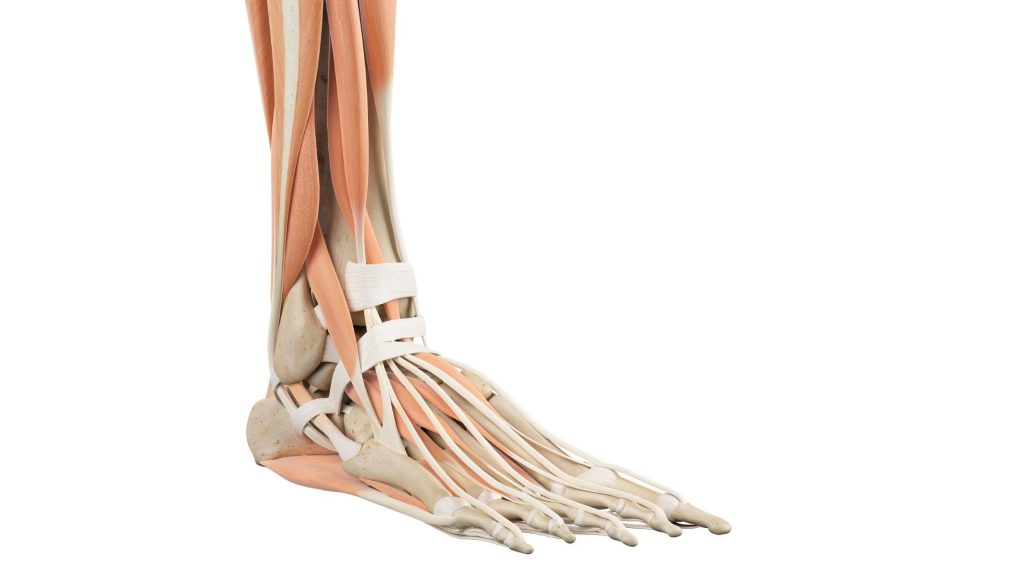
It particularly affects the knee joint but is also seen in the ankle, elbow, shoulder, and hip. This can cause pain, swelling, stiffness, and difficulty moving the affected joint.
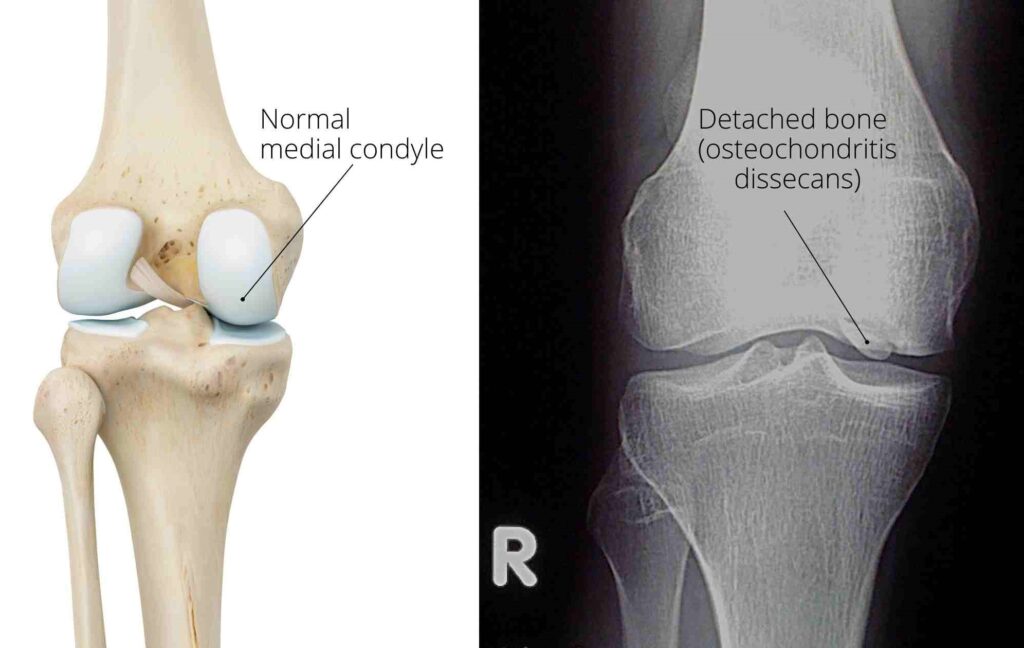
Now, observe the knee joint anatomy figure; on the knee’s inner side is a bony projection called the medial condyle. Due to the disease process, this bony projection undergoes necrosis (death) and becomes soft and detached. All of this results in pain and swelling.
OCD mainly affects young males with a male-to-female ratio of 5:3, mainly affecting the age group of 11 to 20 years3.
The exact cause of osteochondritis dissecans is unclear, but it is thought to be related to a lack of blood flow to the affected area. The disease process makes the part of the bone (inside the joint) soft due to less blood circulation to that part.
A softened bone is vulnerable to deformity and detaches when subjected to pressure or force.
This can be due to repetitive stress on the joint or a traumatic injury. The condition is more common in young athletes who participate in sports that involve running, jumping, or pivoting.
What Causes Osteochondritis Dissecans? The 4 Key Factors
The cause of OCD is considered multifactorial. There may be mechanical, biological, hereditary, and anatomical factors at play. Out of all these, mechanical factors are the most common cause1.
The common mechanical causes include:
- Repetitive stress: Repetitive stress is the most accepted cause, especially in young athletes. A majority of patients (55%–60%) are regularly involved in sports, and repetitive cyclical stress through an athlete’s joint may lead to chondral injury and possible vascular damage, leading to ischemia2.
- Injury
- Impingement against the tibial spine.
- Thrombosis of end artery.
OCD Symptoms: How to Recognize Osteochondritis Dissecans
The symptoms of OCD can be subtle at first and are often mistaken for general growing pains. However, they are typically persistent and directly linked to physical activity.
According to research, the most common presentation is poorly localized, activity-related knee pain, with weight-bearing pain being the predominant symptom in 80% of cases.
- Activity-Related Pain: The most common symptom. The child or adolescent experiences a dull, aching pain in the knee during or after sports, running, or jumping. The pain typically improves with rest.
- Swelling (Joint Effusion): The knee may appear puffy or swollen, often without a specific remembered injury. This is a sign of irritation within the joint.
- Stiffness and Reduced Range of Motion: The knee may not bend or straighten fully. The patient might walk with a slightly bent knee.
- Locking or Catching Sensation: This is a red flag symptom. If the bone fragment has become partially detached (unstable lesion), it can physically catch within the joint, causing the knee to lock in one position. This is often painful.
- Giving Way or Instability: The knee may feel weak or “give out” unexpectedly during weight-bearing activities.
- Limping (Antalgic Gait): To avoid putting full weight on the painful knee, a child may develop a noticeable limp, especially after being active.
When to See a Doctor: If your child experiences persistent knee pain that doesn’t resolve with a few days of rest, especially if accompanied by any swelling, locking, or a limp, it’s essential to consult a healthcare professional for a proper evaluation. Early diagnosis is key to successful treatment.
How is Osteochondritis Dissecans Diagnosed?
If OCD is suspected, a doctor will typically follow a standard process:
- X-rays: This is always the first step. An X-ray can reveal the classic signs of a bone fragment separating. Because OCD is bilateral in up to 30% of cases, X-rays of both knees are often taken.
- MRI (Magnetic Resonance Imaging): This is the most important tool. An MRI not only confirm the diagnosis, but it also helps determine the stability of the fragment. 3If the MRI displays the bright line behind the fragment (indicating fluid) or the presence of cysts it is sign of instability.
Osteochondritis Dissecans Treatment: From Rest to Surgery
The treatment path for OCD isn’t one-size-fits-all. It depends on the patient’s age, symptoms, and the stability of the lesion3
Step 1: Conservative Management (Always the first choice for stable JOCD).
Rest from aggravating activities
According to a 2022 review in Orthopedic Reviews, conservative treatment is the first-line management for juvenile OCD (JOCD) and is often sufficient to ensure healing in patients with open growth plates3.
This often includes sports activities involving jumping, weight-bearing on the knee, running and excessive squatting2.
Immobilization & Physical Therapy:
During adolescence, compliance with activity restrictions may be difficult. In such cases, doctors may immobilise the knee with an off-loading knee brace.
The Role of Physical Therapy in OCD Rehabilitation
In a minor case, the application of ultrasonic therapy may be beneficial. Research suggests that non-weight-bearing exercises can help maintain knee joint flexibility and improve blood circulation. These exercises include swimming and static cycling2.
In the initial phase, knee range of motion exercises and stretching are recommended. Once the full knee range is achieved and partial weight bearing is possible, muscle strengthening exercises and balance training can be added.
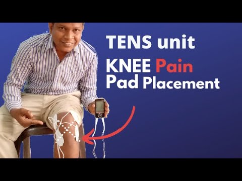
Neuromuscular electrical stimulation, weight-bearing, non-weight-bearing activities, or progressive resistive exercises can facilitate muscle strengthening. Balance activities help restore normal proprioceptive feedback from the knee joint.
In the advanced phase, running and gradually proceeding to impact and sport-specific activities are recommended.
Step 2: Surgical Intervention (If conservative treatment fails)
Surgery is considered if pain persists after 6 months of conservative care or if the lesion shows signs of instability3.
- For Stable Lesions that haven’t healed: Drilling. Surgeons use arthroscopic drilling to create tiny channels in the bone, stimulating blood flow and healing. This can be highly effective3.
- For Unstable Lesions: Fixation. The loose fragment is reattached using bioabsorbable pins or metal screws.
- For Severely Damaged Lesions: Salvage Procedures. If the fragment is too damaged to be reattached, procedures like microfracture, osteochondral grafting (OATS), or autologous chondrocyte implantation (ACI) are used to repair the defect3.
What is the Prognosis for OCD?
There are several factors that affect the likelihood of a full recovery3:
- Age and Skeletal Maturity: Children and adolescents with open growth plates (Juvenile OCD) have a much better prognosis than adults (Adult OCD). Within the juvenile group, children under 12 have the best outcomes.
- Lesion Stability: Stable lesions (where the bone and cartilage fragment is still in place) heal well with conservative care. Unstable or detached fragments require surgery.
- Lesion Size and Location: Smaller lesions (<2 cm²) generally do better. Lesions on the medial femoral condyle have a good prognosis, while those on the patella (kneecap) are more challenging.
FAQ
The author is a physiotherapist who has been practising for the last 17 years. He holds a Bachelor's in Physiotherapy (BPT) from SVNIRTAR (Swami Vivekananda National Institute of Rehabilitation and Research), one of the prestigious physiotherapy schools in India.
Whatever he learns dealing with his patient, he shares it with the world through blogs and e-books. He also owns a YouTube channel, "Sunit Physiotherapist" with over 8 lakh active subscribers. Here, he shares everything he gets to learn serving the patient.


Pingback: Rheumatoid arthritis vs osteoarthritis: which arthritis is worse? : Physiosunit
This is a fundamental principle on how your body is designed to function. If you have pain in your knees your body compensates in the hips, lower back, upper back, neck, shoulders, and arms. kniepijn
I hope you will share such type of impressive contents again with us so that we can utilize it and get more advantage. joint supplements,
Nice post, Thank you for sharing valuable information. I enjoyed reading this post. The whole blog is very nice found some good stuff and good information here Thanks for sharing…Also visit my page.
Premium Folding Wheelchair