Last Updated on October 15, 2025 by Sunit. S. Ekka

What Will You Learn?
- The main causes and mechanisms behind mid-shaft humerus fractures
- How to classify and recognize different types of humerus fractures
- Key clinical signs, symptoms, and how to assess for complications like nerve injury
- Essential treatment options: when to use conservative or surgical approaches
- The most common complications and how to prevent them
- Simple, effective rehabilitation exercises to regain strength and restore movement in your shoulder, elbow, and wrist
A mid-shaft humerus fracture is a common injury resulting from falls, direct trauma, or sports accidents. Understanding the mechanism of injury, types and classification, clinical features, investigations, and complications is important.
In this comprehensive guide, we not only explain the basics of mid-shaft humerus fractures but also provide a step-by-step, evidence-based approach to physiotherapy management. You’ll learn the rehabilitation exercises for regaining shoulder, elbow, and wrist mobility after a plaster cast is removed.
- 8 Best Humerus Fracture Exercises to Regain Elbow Mobility (At Home)
- Exercise 1: Wrist Flexion and Extension for Humerus Fracture Recovery
- Exercise 2: Wrist Circles to Improve Flexibility After a Broken Arm
- Exercise 3: Wrist Flexor/Extensor Stretch (Step-by-Step Guide)
- Exercise 4: Forearm Rotation (Supination/Pronation) to Restore Movement
- Exercise 5: Grip Strengthening with a Gel Ball (Prevent Weakness)
- Exercise 6: Shoulder Elevation to Prevent Stiffness After Immobilization
- Phase 2: Post-Cast Rehab (6 Weeks After Plaster Removal)
- Exercise 7: Elbow Flexor/Extensor Stretch (Reduce Post-Fracture Tightness)
- Exercise 8: Dumbbell Strengthening for Long-Term Humerus Fracture Recovery
- Humerus Shaft Fracture: Types (Spiral, Oblique, Comminuted) Explained
- Broken Arm Treatment: Surgical vs. Non-Surgical Options
- Common Complications After Humerus Shaft Fracture
- Key Takeaways for Humerus Fracture Rehabilitation
- FAQ
8 Best Humerus Fracture Exercises to Regain Elbow Mobility (At Home)
After a fractured humerus is surgically managed, the patient must be immobilised for a certain number of weeks. It is during this immobilisation period that most complications arise.
The most common complication is stiffness in the elbow joint. However, stiffness can also affect the wrist and shoulder joints.
To reduce the risk of complications, it is recommended to begin physiotherapy as soon as the plaster cast is removed and the initial pain and swelling have subsided.
The exercises we will cover are designed to strengthen the muscles of the arm and forearm and help regain the range of motion in the elbow, wrist, and shoulder joints. So, let us start with wrist flexion-extension exercises.
Exercise 1: Wrist Flexion and Extension for Humerus Fracture Recovery

Let’s begin by discussing the elbow joint, which is often the most affected joint. The elbow joint can perform four movements: flexion, extension, pronation, and supination. Initially, we will aim to restore elbow flexion and extension motion.
- To do this, find a comfortable sitting position in front of a table and place the affected arm on the table.
- Keep the arm flat on the table to provide a stable base for elbow exercises. In this starting position, fully straighten the elbow, known as elbow extension.
- After fully extending the elbow, slowly bend it, known as elbow flexion.
- Repeat this alternating flexion-extension movement at least 15 to 20 times per session.
Exercise 2: Wrist Circles to Improve Flexibility After a Broken Arm

- To begin this exercise, make sure you are in a comfortable and relaxed position, similar to the previous exercise.
- Once you are comfortable, focus on rotating your wrist joint. Start by rotating it in a clockwise direction around 15 to 20 times, ensuring that you move your wrist in a smooth and controlled manner. You should feel a gentle stretch in your wrist as you rotate it.
- After you have completed the clockwise rotation, switch to rotating your wrist in an anticlockwise direction for another 15 to 20 times. Remember to keep your movements smooth and controlled.
- You can repeat this exercise as many times as you like, depending on your comfort level.
- This exercise is great for improving flexibility and mobility in your wrist joint and can help reduce stiffness and discomfort.
Exercise 3: Wrist Flexor/Extensor Stretch (Step-by-Step Guide)

- To stretch the wrist, our starting position remains the same.
- To begin, we must use our opposite hand and gently apply pressure at the end range of wrist flexion and extension.
- For instance, if we want to stretch the flexors, we should slightly extend the wrist and then use our other hand to apply pressure over the palm. This will provide the necessary tension to the flexor muscles.
- We should maintain this position for at least 30 seconds to ensure that the muscles get correctly stretched.
- We can then repeat the same process by extending the wrist to stretch the flexor group of muscles.
- With proper execution, this exercise can help improve the flexibility of our wrist muscles and prevent injuries.
Exercise 4: Forearm Rotation (Supination/Pronation) to Restore Movement

When it comes to exercises for a humerus shaft fracture, it’s important not to overlook forearm pronation and supination, which are necessary elbow joint motions.
- To perform this exercise, start by stabilizing your forearm and keeping it flat on a table, as shown in the figure.
- From this position, rotate your forearm clockwise and anticlockwise. Clockwise rotation is called supination, while anticlockwise rotation is called forearm pronation. Initially, you can do this exercise without holding anything.
- Still, as you become more comfortable with it, it is recommended to hold a cane or a lightweight rod to make the exercise more effective. The momentum created by the cane will help you move the forearm in both directions with greater ease.
Also Read: Supracondylar Fracture of Humerus: Physiotherapy management
Exercise 5: Grip Strengthening with a Gel Ball (Prevent Weakness)

Long-term immobilisation can cause grip weakness, making it difficult to hold objects such as a glass of water. Strengthening the grip is crucial, along with continuing other recommended exercises.
To improve grip strength, a silicone gel ball can be used by squeezing it as many times as possible. This simple yet effective exercise can be performed frequently.
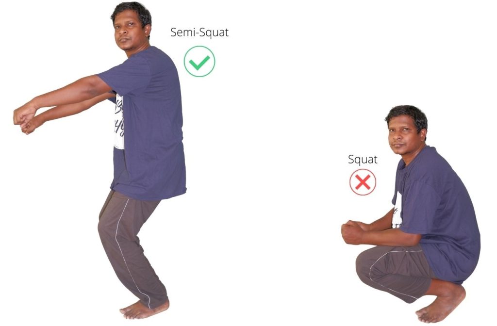
Exercise 6: Shoulder Elevation to Prevent Stiffness After Immobilization

Often, shoulder joint stiffness is ignored in such conditions. Shoulder stiffness can become problematic if not taken care of.
We have to start shoulder exercises along with other shoulder stiffness exercises. So, this shoulder elevation exercise is a very simple, basic exercise. For this:
- stand straight and srasp your hand n front of you.
- Now, elevate it to the maximum point where you can take it.
- Elevate it and then lower it down, and then repeat the same process 15 to 20 times in a single session.
- Twice daily is recommended.
Phase 2: Post-Cast Rehab (6 Weeks After Plaster Removal)
After removing the plaster (or another sling), the most important complaint is pain in the shoulder and elbow. The person has difficulty keeping the elbow straight, including the shoulder joint restriction.
For elbow joint restriction, do the following exercises:
Exercise 7: Elbow Flexor/Extensor Stretch (Reduce Post-Fracture Tightness)

Once again, it is a very important stretching exercise for managing the shaft of a humerus fracture. This is a stretching exercise for the elbow flexor and extensor groups of muscles.
Flexor muscles are present on the front of the elbow, and their action brings flexion movement to the elbow joint. Extensor muscle brings extension/ straightening of the elbow joint and is present on the back of the elbow.
- Let us start with stretching of the elbow flexors; first, straighten your elbow to the maximum.
- In this extended elbow position, put slight overpressure with the opposite hand till you feel a comfortable stretch on the front of the elbow.
- Hold this stretched position for a minimum of 30 seconds. Repeat the same process to stretch the elbow extensors, but this time by bending the elbow.
- Do it 3 to 5 times in a single session.
Exercise 8: Dumbbell Strengthening for Long-Term Humerus Fracture Recovery

To perform this exercise, it is recommended to use 1.5 to 2 kg dumbbells. However, it’s important to note that this exercise should only be attempted if you have gained sufficient grip strength to hold the dumbbell firmly.
- To begin, stand straight and hold the dumbbell with the hand on the affected side. When holding the dumbbell, allow the weight to exert a gentle stretch on the elbow joint.
- It is suggested to hold the dumbbell for at least one minute, but you can hold it for up to five minutes based on your comfort level.
- Over time, you can gradually increase the weight of the dumbbells up to 5 kg as you become more comfortable with the exercise.
- Remember to take it slow and steady, and always listen to your body’s signals to avoid discomfort or injury.
Humerus Shaft Fracture: Types (Spiral, Oblique, Comminuted) Explained
When we suffer a fracture in our arm, it is typically a fracture of the humerus bone. Although this type of fracture is more commonly seen in adults, it can also occur in children, albeit in rare cases.
The fracture in the shaft of the humerus can be classified into three types.
In our previous post titled “3 Fracture Humerus Classification, Its Sub-Type Simplified,” we explained that our arm can fracture in three different sites: first, on the head or neck of the humerus; second, on the shaft of the humerus; and third, on the epicondyles of the humerus.
However, in order to better understand the concept of a fracture, we must first have a basic understanding of its related anatomy.

By Bduttabaruah – https://commons.wikimedia.org/wiki/File:HumerusBack.png, CC0,
The humerus bone is the only bone in our arm. It is a single, long bone with three parts: the head, the epicondyles, and the shaft. The shaft is cylindrical, slightly flattened on the upper part, and conical on the lower part.
It is surrounded by bulky muscles such as the biceps, triceps, and deltoid.
The humerus bone has a rich blood supply, which means that any fracture on the shaft heals readily. The most common site of fracture is at the junction of the upper and lower parts of the bone.
Fractures can occur due to direct injury or assault on the arm, or sudden, opposite movement of the arm against the violent contraction of the muscle.
Depending on the fracture pattern, it can be categorised into three types: spiral, oblique, and comminuted.
Spiral fracture

As the term suggests, the fracture occurs in a spiral pattern. This occurs due to strong rotational movement creating a twisting force on the humerus.
Oblique / transverse fracture
The fracture makes a transverse pattern, as shown in the figure. This fracture occurs due to the strong angular force exerted on the middle area of the shaft.

Comminuted fracture

When there is direct injury or assault on the arm, the bone may break into multiple pieces. The fracture becomes fragmented.
Broken Arm Treatment: Surgical vs. Non-Surgical Options
Management depends upon whether the fracture is stable or unstable. The stable fracture can be managed conservatively, but the unstable fracture needs surgical intervention.
Conservative management:
The displaced fracture, where fracture segments get displaced from each other, is first re-aligned by the surgeon. The entire process is carried out under the effect of anaesthesia.
After the fracture reduction, the immobilisation of the arm is done for 6 to 8 weeks using one of the following ways. U slab Hanging cast. Chest arm bandage.
Surgical management:
If the fracture is unstable or if it is associated with multiple other bone fractures, then surgery is considered. The following is the surgical process for such fractures.
- Intra-medullary nailing.
- Screw and plate.
- After the operation, the arm is immobilised using the sling support (or shoulder spica) for six weeks.
Common Complications After Humerus Shaft Fracture
- Malunion: Some amount of malunion is accepted.
- Non-union: Non-union occurs due to the inadequate reduction/ distraction or immobilisation
- Radial nerve injury:
1) Radial nerve injury can occur at the time of fracture, during fracture reduction or after healing (nerve gets entrapped in the callus).
2) Usually, nerve injury is a neuropraxia type, which heals within 2-3 weeks.
3) Radial nerve injury can cause wrist drop.
Key Takeaways for Humerus Fracture Rehabilitation
Sometimes, this fracture is associated with radial nerve injury, resulting in wrist drop. In some rare cases, there may be malunion delayed / non-union.
This is a special situation that needs a different treatment approach altogether, and it is beyond the scope of this post. In my practice, I have found that elbow restriction is the most common complaint after the removal of plaster. So, proper exercise is very crucial.
Keep Reading: Supracondylar Fracture of Humerus: Physiotherapy management
FAQ
The author is a physiotherapist who has been practising for the last 17 years. He holds a Bachelor's in Physiotherapy (BPT) from SVNIRTAR (Swami Vivekananda National Institute of Rehabilitation and Research), one of the prestigious physiotherapy schools in India.
Whatever he learns dealing with his patient, he shares it with the world through blogs and e-books. He also owns a YouTube channel, "Sunit Physiotherapist" with over 8 lakh active subscribers. Here, he shares everything he gets to learn serving the patient.


Pingback: What is frozen shoulder: 3 Stage & Physiotherapy to Ease Pain - Physiosunit
Pingback: Fracture humerus classification and causes - Physiosunit
Pingback: How to Loosen Tight Shoulders? 7 Exercises fix tight shoulders - Physiosunit
Very sorry. I have rectified it
it is 2018 and there is no rectification yet if you have no intention to do so just take it off
Thank you for pointing it out. will rectify it soon
Where did you get this annotated humerus picture from??
Greater Tubercle – NOT greater trochanter
Medial and Lateral Epicondyle – NOT malleoli
Trochlea – NOT condylar