Last updated on February 15th, 2024 at 07:07 pm

mage courtesy of kdshutterman at FreeDigitalPhotos.net
- Osteochondritis dissecans of the knee is a condition that affects the cartilage and bone in the knee joint.
- This type of joint disorder typically occurs in children and young adults.
- The condition can cause pain, swelling, and limited range of motion in the knee joint.
- Osteochondritis dissecans can be caused by a variety of factors, including trauma, repetitive stress, and genetic predisposition.
- Treatment of the condition may involve rest, physical therapy, and, in some cases, surgery.
If your child complains of knee pain after physical exertion or playing outdoor games for some time, it may be due to osteochondritis dissecans. Along with pain, they will also complain of knee swelling and sometimes difficulty in moving the knee joint with limping. This condition is normally seen in skeletally immature persons such as children, but sometimes, it is also reported in young adults1.
This article will teach what causes osteochondritis dissecans in children and the available treatment options. We will also learn if physiotherapy can help them ease their pain.
What is osteochondritis dissecans?
Osteochondritis dissecans is a medical condition that affects the joints where a piece of bone and cartilage in the joint becomes loose and separates from the rest of the bone. It particularly affects the knee joint but is also seen in the ankle, elbow, shoulder, and hip. This can cause pain, swelling, stiffness, and difficulty moving the affected joint.
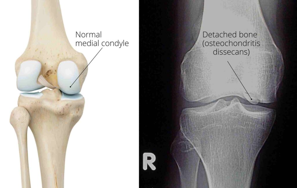
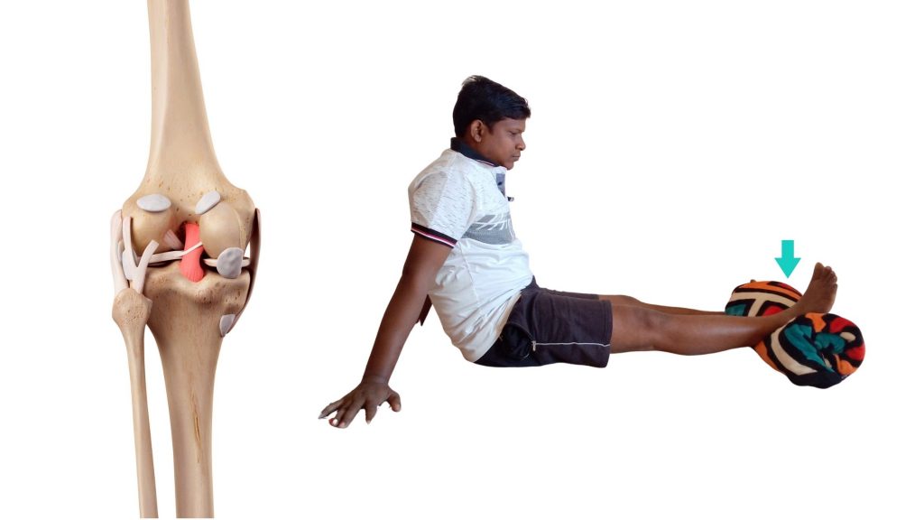
Now, observe the knee joint anatomy figure; on the knee’s inner side is a bony projection called the medial condyle. Due to the disease process, this bony projection gets necrosis (dead) and becomes soft and detached. All of this results in pain and swelling.
The exact cause of osteochondritis dissecans is unclear, but it is thought to be related to a lack of blood flow to the affected area. The disease process makes the part of the bone (inside the joint) soft due to less blood circulation to that part. A softened bone is vulnerable to deformity and detaches when subjected to pressure or force.
This can be due to repetitive stress on the joint or a traumatic injury. The condition is more common in young athletes who participate in sports that involve running, jumping, or pivoting.
Factors that causes osteochondritis dissecans
The exact causative factors for the condition are not yet clear. There may be mechanical, biological, hereditary, and anatomical factors at play. Out of all these, mechanical factors are the most common cause1, which can be a single major trauma or a repetitive micro-trauma/stress.
Repetitive stress is the most accepted cause, especially in young athlete. A majority of patients (55%–60%) are regularly involved in sports, and repetitive cyclical stress through an athlete’s joint may lead to chondral injury and possible vascular damage, leading to ischemia2.
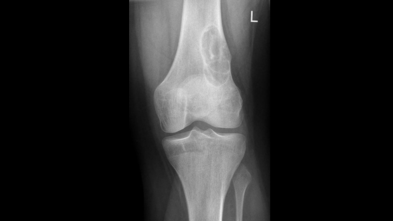
Localised avascular necrosis of articular cartilage of medial femoral condyle resulting in softening. In some extreme cases, detachment of the bony projection may happen.
- Injury.
- Impingement against the tibial spine.
- Thrombosis of end artery.
Clinical feature
- Pain on exertion. The child will complain of severe pain after physical activity.
- Joint effusion present. Painful swelling and inflammation of the knee joint.
- The loose body inside the joint results in locking and restriction of the terminal extension of the knee.
- Joint effusion and restriction of range of motion.
Treatment
Rest from aggravating activities
Conservative management is the first choice of treatment for osteochondritis dissecans. The sufferer is immediately restricted from activities that aggravate the pain. This often includes sports activities involving jumping, weight-bearing on the knee, running and excessive squatting2.
During adolescence, compliance with activity restrictions may be difficult. In such cases, doctors may immobilize the knee with an off-loading knee brace.
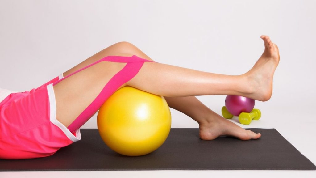
Physical therapy and exercises
In a minor case, the application of ultrasonic therapy may be beneficial. Research suggests that non-weight-bearing exercises can help maintain knee joint flexibility and improve blood circulation. These exercises include swimming and static cycling2.
In the initial phase, knee range of motion exercises and stretching are recommended. Once the full knee range is achieved and partial weight bearing is possible, muscle strengthening exercises and balance training can be added. Neuromuscular electrical stimulation, weight-bearing, non-weight-bearing activities, or progressive resistive exercises can facilitate muscle strengthening. Balance activities help restore normal proprioceptive feedback from the knee joint. In the advanced phase, running and gradually proceeding to impact and sport-specific activities are recommended.
Surgical management
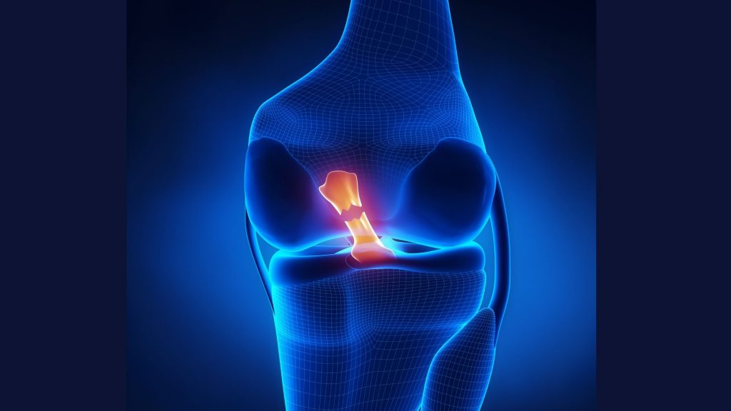
In a late case or severe case where there is already bony projection is detached, surgery is the only option left. The surgical process involves removal of the loose body form the joint.
The author is a physiotherapist who has been practising for the last 17 years. He holds a Bachelor's in Physiotherapy (BPT) from SVNIRTAR (Swami Vivekananda National Institute of Rehabilitation and Research), one of the prestigious physiotherapy schools in India.
Whatever he learns dealing with his patient, he shares it with the world through blogs and e-books. He also owns a YouTube channel, "Sunit Physiotherapist" with over 8 lakh active subscribers. Here, he shares everything he gets to learn serving the patient.

Pingback: Rheumatoid arthritis vs osteoarthritis: which arthritis is worse? : Physiosunit
This is a fundamental principle on how your body is designed to function. If you have pain in your knees your body compensates in the hips, lower back, upper back, neck, shoulders, and arms. kniepijn
I hope you will share such type of impressive contents again with us so that we can utilize it and get more advantage. joint supplements,
Nice post, Thank you for sharing valuable information. I enjoyed reading this post. The whole blog is very nice found some good stuff and good information here Thanks for sharing…Also visit my page.
Premium Folding Wheelchair