Last updated on May 24th, 2024 at 05:09 pm
The heart is a pumping machine that collects impure blood from our body and pumps it to the lungs for purification. The pure blood is again collected in the heart, which is once again pumped to different parts, muscles, cells, and tissues of our body.
All these events happen smoothly in a cycle known as the cardiac cycle. In this article, we will try to learn the cardiac cycle and what those curves and plateaus denote in a cardiac cycle.
Cardiac cycle explained
Our heart consists of four chambers: two on the left and two on the right. We call them the Right atrium, Right ventricle, Left atrium, or Left ventricle (see figure above).
Each chamber has an opening to allow blood to flow inside, and each opening has a lid that checks blood flow. We call this lid a valve. Our heart has four valves.
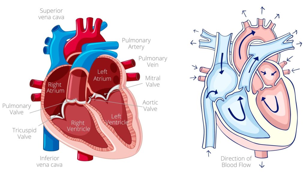
- Between the left atrium and left ventricle is the mitral valve.
- The tricuspid valve is on the right side, between the right atrium and right ventricle.
- The right ventricle opens in the pulmonary artery, and a pulmonary valve exists between them.
- Similarly, the left ventricle opens in the arch of the aorta, and the aortic valve lies between them.
Cardiac cycle phases
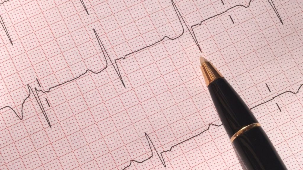
The heart is the most hardworking organ, supplying blood to every corner through the circulatory system. We can electronically measure this cardiac cycle; in science, we call this process measuring ECG (Electro Cardio Gram).
Broadly, the cardiac cycle passes through two phases:
- Diastole: It is a period of relaxation of the heart chamber when the heart is refilled.
- Systole: The period of contraction of the heart chamber, and blood is pumped out of the heart.
These two phases are subdivided into seven phases. These are:
- Atrial systole.
- Isovolumetric contraction.
- Rapid ventricular ejection.
- Reduced ventricular ejection.
- Isovolumetric relaxation.
- Rapid ventricular filling.
- Reduced ventricular filling.
Don’t get confused by all these terms. We will discuss them in detail, and I will try to make it as simple as possible. At the end of this subheading, you will find a table of all phases and their events. So, let us start one by one.
Also read: Which organ is called second heart of the body & why?
Phase 1) Atrial systole
Atrial systole is the phase of atrial contraction. During this phase, the atrium contracts to pump blood into the ventricles. However, only 20% of ventricle filling occurs by atrial systole. The rest of the 80% of ventricular filling has been done passively, even before the onset of atrial contraction. This active filling of the ventricles becomes valuable during physical activity.
When the pressure in the atrium increases, blood rushes into the ventricles through the opened mitral valve. During left atrium contraction, pressure and volume are transferred into the left ventricle through the opened mitral valve.
Remember, the aortic valve is closed because the pressure in the aorta is greater than the pressure in the left ventricle at this moment.
To understand it clearly, let us once again go back to the above figure and observe the arrow. As you can see, the arrow shows the direction of blood flow from the superior and inferior vena cava, which supplies blood into the right Atrium, and the arrow on the pulmonary Veins shows the direction of blood flow to the left Atrium.
Both the atrioventricular valves are open; blood from the left and right Atrium goes into the left and right ventricles.
Phase 2) Isovolumetric contraction
It is the initial phase of the ventricle systole, which means the ventricles are in a state of contraction. In this phase, the contraction ventricle starts, and the pressure inside the chamber starts to build.
However, initially, the pressure inside the ventricle is not sufficient to push open the semilunar valve. Soon, however, this initial pressure rises above the pressure of the atrium, resulting in the closure of the atrioventricular.
Phase 3) Rapid ventricular ejection
This is the second phase of ventricular systole, and pressure inside the ventricles slowly increases. As the pressure builds up, it soon reaches a point where it pushes open the arctic and pulmonary valves and pumps out blood from the heart.
Since blood is ejaculated rapidly from the ventricle, this phase is called the Rapid ventricular ejection phase. During this phase, 70% of the blood is empty.
Phase 4) Reduced ventricular ejection
Contrary to the above, it is a phase of reduced ventricular ejection. It is the last phase of the ventricle systole, during which the pressure inside the ventricles decreases, and the rest 30% of the blood is emptied.
Phase 5) Isovolumetric relaxation phase
A sub-phase of ventricle diastole occurs during which the ventricle comes to a state of relaxation, and the isovolumetric relaxation phase is the early phase of ventricle diastole.
As the ventricle muscles relax, the pressure inside the ventricle dips. When it decreases below the pulmonary trunk and aorta pressure, the blood rushes back towards the heart. The backward flow of blood causes the closure of the semilunar valve.
During this phase, the atrioventricular valves remain closed, and the volume of the blood in the ventricle does not change, which is why it is called an isovolumetric relaxation phase.
Phase 6) Rapid ventricular filling phase
The rapid ventricular filling phase is the second phase of ventricle diastole, during which the ventricle muscles relax further, and pressure decreases further. Eventually, the pressure inside the ventricle drops to a point where it becomes lower than the pressure inside the atria.
As a result, blood from the atrium pushes open the atrioventricular valve, and blood rushes into the ventricles. This happens rapidly, so it is called a Rapid ventricle filling phase.
Phase 7) Reduced ventricular filling
During the reduced ventricle filling phase, which is also known as diastasis, the ventricles continue to fill slowly. This phase represents the longest part of the cardiac cycle and occurs after the rapid filling phase.
During diastasis, the heart muscle is in a state of relaxation, allowing for a gradual and steady filling of the ventricles with blood from the atria. This phase is crucial for ensuring that the ventricles are adequately filled before the next contraction, which is essential for maintaining an efficient cardiac output.
Table of phases of the cardiac cycle
| Phases of cardiac cycle | Semilunar valve | AV valve | |
| 1 | Atrial systole | closed | open |
| 2 | Isovolumetric contraction | closed | closed |
| 3 | Rapid ventricular ejection | open | closed |
| 4 | Reduced ventricular ejection | open | closed |
| 5 | Isovolumetric relaxation phase | closed | closed |
| 6 | Rapid ventricular filling phase | closed | open |
| 7 | Reduced ventricular filling | closed | open |
Cardiac cycle ECG
We can study these cardiac cycle phases by analysing the heart’s electrical activity. This process has been termed an electrocardiogram (ECG/ EKG). I have covered this topic extensively in one of my articles How to read ECG: Electrocardiogram Simplified.
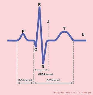
Here’s a summary of what an ECG wave contains.
P stands for atrial systole.
Q, R, S complex stands for ventricle systole.
T, U stand for diastole.
Keep Reading: Difference Between Stroke and Heart Attack
The author is a physiotherapist who has been practising for the last 17 years. He holds a Bachelor's in Physiotherapy (BPT) from SVNIRTAR (Swami Vivekananda National Institute of Rehabilitation and Research), one of the prestigious physiotherapy schools in India.
Whatever he learns dealing with his patient, he shares it with the world through blogs and e-books. He also owns a YouTube channel, "Sunit Physiotherapist" with over 8 lakh active subscribers. Here, he shares everything he gets to learn serving the patient.


Thank you
Nice post, Thank you for sharing valuable information. I enjoyed reading this post. The whole blog is very nice found some good stuff and good information here Thanks for sharing…Also visit my page.
Premium Folding Wheelchair