Welcome, students! In this first chapter of our electrotherapy series, we will explore the foundational concepts of physics that underpin the electrical and electromagnetic equipment used in physiotherapy.
The knowledge shared here is derived from Clayton’s textbook on electrotherapy (now continued as Watson’s Electrotherapy: Evidence-Based Practice).1 The aim is to provide a simplified and clear introduction so you can confidently understand how therapeutic modalities interact with the body at a basic physical level.
This session covers the core ideas you need as you begin your journey in electrotherapy. We will examine the structure of the atom and how electron transitions produce electromagnetic radiation — the same radiation that forms the basis of many electrotherapy treatments.
Topics Covered in This Session
Atoms and Atomic Structure
Let us begin by understanding the atom and its structure.
Atoms are the basic building blocks of all matter. Just as cells form the foundation of living tissue in biology, atoms form the foundation of the physical world around us — including the tissues and fluids of the human body that we treat as physiotherapists.
What is an atom exactly?
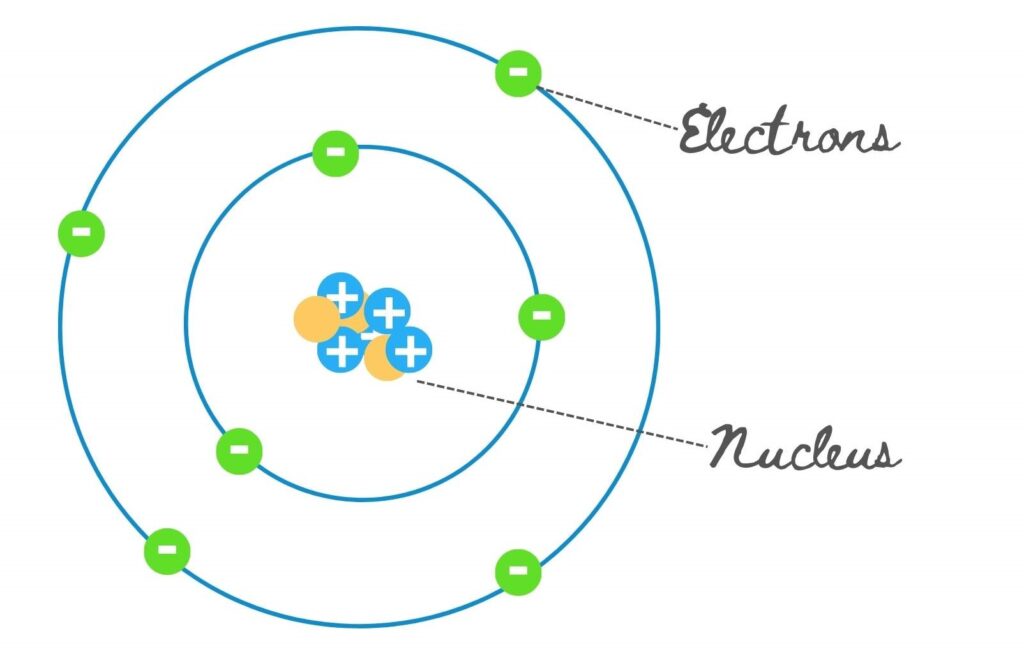
In the simplified (Bohr) model commonly used for introductory teaching, an atom consists of a central nucleus surrounded by electrons that occupy definite energy levels or orbits. Every electron moves within a specific energy level. (Note: modern quantum mechanics describes electron behaviour in terms of probability clouds rather than fixed planetary-style orbits, but the Bohr model remains a useful teaching tool for understanding energy transitions.)
A helpful analogy is the solar system: the sun represents the nucleus at the centre, while the planets represent electrons revolving around it in fixed paths.
Nucleus and its Components
The nucleus of an atom contains protons and neutrons. Protons are positively charged particles (often denoted “P” for positive).
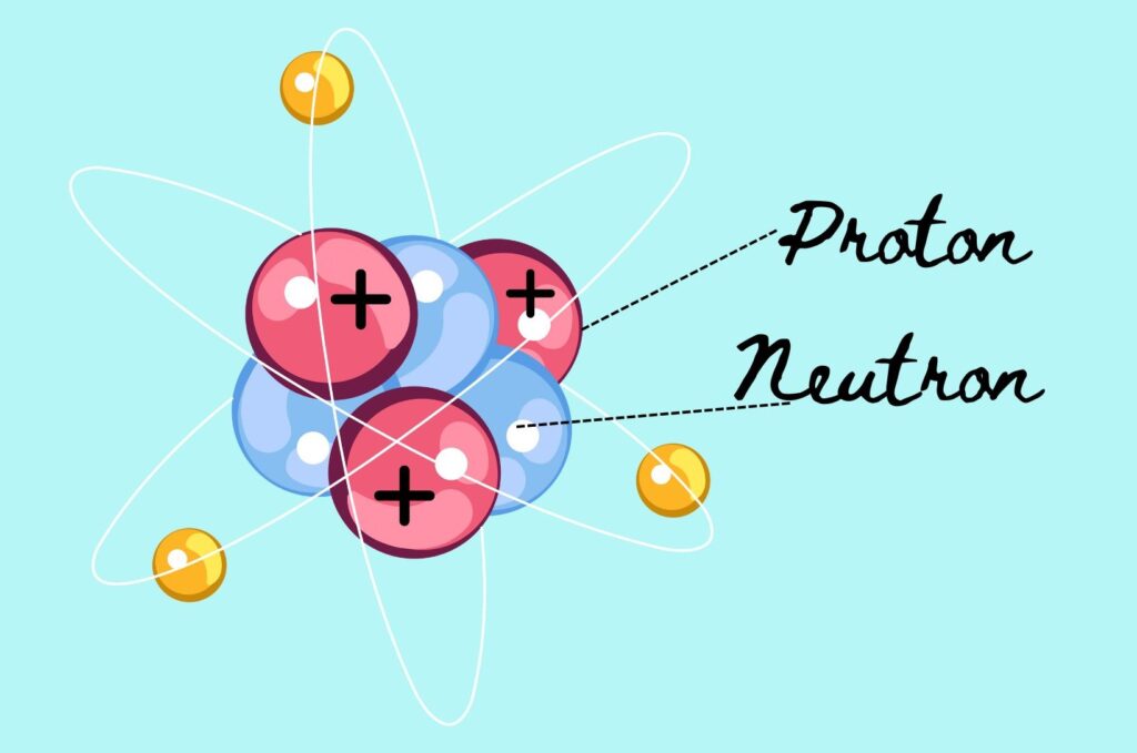
Neutrons are neutral particles (symbolised “N”). Strong nuclear forces hold these protons and neutrons tightly together, keeping the nucleus compact and stable.
Negatively charged electrons surround the nucleus. They occupy energy levels (orbits) and create a cloud of negative charge that largely determines the size of the atom.
Protons are relatively large nuclear particles with a positive charge, while neutrons have a mass almost equal to protons but carry no charge. The balance between these particles is critical for the stability of the atom.
Understanding these components is essential because the movement of electrons between energy levels is what produces electromagnetic radiation — the physical basis of many electrotherapy modalities used in physiotherapy practice.
Arrangement in the Periodic Table
The arrangement of subatomic particles determines the chemical properties of an element. Protons, neutrons and electrons together decide the characteristics of each element.
The number of protons in the nucleus (the atomic number) uniquely defines an element and determines its position in the periodic table. Each element has its own place based on this proton count.
Electrons are responsible for chemical bonding. When atoms interact, their outer electrons form bonds that create molecules and compounds.
A classic example is water (H2O): two hydrogen atoms and one oxygen atom share electrons to form a stable molecule. Another example is common table salt (NaCl), formed when sodium and chlorine atoms transfer electrons and bond ionically.
These interactions between atoms are fundamental to the chemistry of body tissues and to the physical principles that govern how therapeutic agents interact with those tissues.
Introduction to Electromagnetic Radiation
Electrons surround the atom’s nucleus in defined energy levels. Electromagnetic radiation is produced when an electron absorbs energy, jumps to a higher energy level, and then returns to a lower level, releasing the excess energy as a photon (an electromagnetic wave).
The type of electromagnetic wave produced depends on the size of the energy transition. Larger energy gaps produce higher-frequency radiation; smaller gaps produce lower-frequency radiation.
A familiar example is tungsten, the metal used in traditional light-bulb filaments. When the filament is heated:
- At lower temperatures it first emits infrared radiation (felt as heat).
- As temperature rises and energy gaps increase, it begins to emit visible light — first red, then yellow, and finally white.
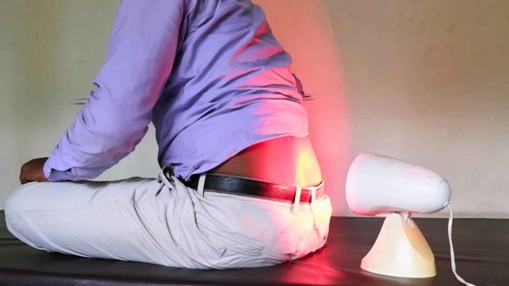
This same principle of electron energy transitions underlies many electrotherapy modalities. Infrared lamps deliver thermal radiation for superficial heating. Ultraviolet radiation is used for certain dermatological and wound-care applications.
Shortwave and microwave diathermy rely on electromagnetic energy at specific frequencies to produce deep tissue heating. Understanding atomic structure and electron transitions therefore helps the physiotherapist appreciate why different modalities produce different physiological effects and how to apply them safely and effectively.
Key Takeaways
- Atoms consist of a nucleus (protons + neutrons) and surrounding electrons.
- The number of protons defines the element and its place in the periodic table.
- Electrons occupy energy levels; transitions between levels produce electromagnetic radiation.
- This radiation is the physical basis of modalities such as infrared therapy, ultraviolet therapy and diathermy.
- A solid grasp of atomic structure electrotherapy concepts forms the foundation for understanding how therapeutic equipment works and how it interacts with body tissues.
What’s Next?
In the following sessions we will build on these fundamentals and examine specific forms of electromagnetic energy used in clinical practice, starting with the properties and therapeutic applications of infrared radiation. You may also explore related topics such as current electricity and electromotive force and therapeutic ultrasound as you progress through the electrotherapy series.
For a visual walkthrough of the same material, watch the companion video lesson.
References
- Watson T, editor. Electrotherapy: Evidence-Based Practice (formerly Clayton’s Electrotherapy). 12th ed. Edinburgh: Elsevier; 2008.
- OpenStax / Physics LibreTexts. Atomic Spectra and X-rays. Available from: phys.libretexts.org (standard physics explanation of electron energy-level transitions and photon emission).
- Tsai SR, Hamblin MR. Biological effects and medical applications of infrared radiation. J Photochem Photobiol B. 2017;170:197-207. doi:10.1016/j.jphotobiol.2017.04.014. PMID: 28441605. Free full text: PMC5505738.
FAQ

The author is a physiotherapist who has been practising for the last 17 years. He holds a Bachelor's in Physiotherapy (BPT) from SVNIRTAR (Swami Vivekananda National Institute of Rehabilitation and Research), one of the prestigious physiotherapy schools in India.
Whatever he learns dealing with his patient, he shares it with the world through blogs and e-books. He also owns a YouTube channel, "Sunit Physiotherapist" with over 8 lakh active subscribers. Here, he shares everything he gets to learn serving the patient.