
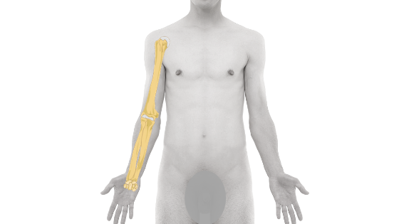
The human arm is built on a framework of three key bones: one in the upper arm (the humerus) and two in the forearm (the radius and ulna). Understanding their structure is essential to knowing how your arm moves and what happens when a common injury, like a fracture, occurs. This guide breaks down the anatomy and injuries of each bone.
The human arm has three bones — the humerus in the upper arm, and the radius and ulna in the forearm.
The human arm, or simply the human upper limb, has an upper arm and forearm. The upper arm bone consists of a single long and strong bone, and there are two bones in the forearm. So, the human arm consists of three long bones in total.
We all know the function of our arm and hand; in this article, we will discuss what these three bones are. What we call it and its few anatomical features. We will also cover the possible fractures these bones often encounter.
So, without delay, let’s get started.
The Three Arm Bones: Humerus, Radius, and Ulna
The bone of the upper arm is known as the humerus bone; it is comparatively stronger than the bones of the forearm. Our forearm consists of two bones. These are the radius and ulna bones. Let us start with the humerus bone.
Humerus Bone: Anatomy of the Upper Arm
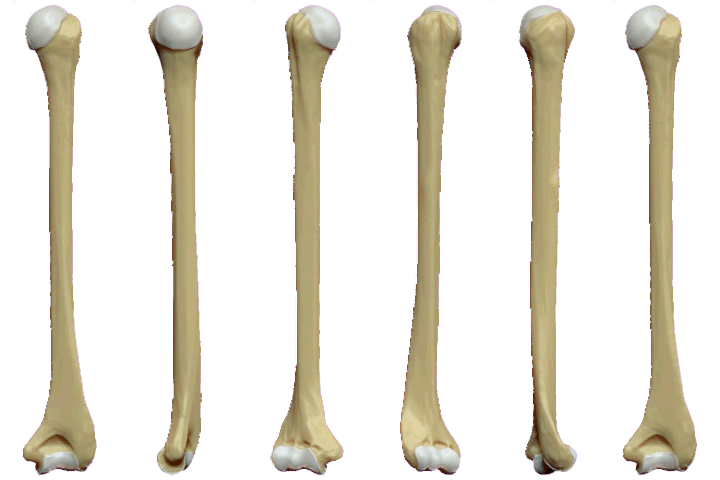
As you can see in the figure, the humerus bone is a thick, long bone with a rounded upper end. Its lower end is slightly triangular and flat. We call the upper end as head.
The head is hemispherical and fits into the outer end of the scapula to make the shoulder joint. This hemispherical shape of the head of the humerus gives it a great degree of freedom at the shoulder joint.
Just move your shoulder and observe that we can do all the following movements, like:
- Shoulder elevation,
- Extension,
- External rotation of the shoulder
- Shoulder internal rotation,
- Abduction,
- Adduction,
- Shoulder circumduction
Just below the head of the humerus, the bone takes a long and thick cylindrical shape that forms the shaft of the humerus. On its upper end, the shaft is a bit circular in cross-section it gets flat at the lower end.
At the end of the flat and triangular lower end of the shaft is an articular surface known as a condyle. This articular surface, together with the upper end of the bones of the forearm, makes an elbow joint. When the arm is fully extended, the alignment of the humerus and forearm forms the carrying angle of the elbow.
Common Humerus Fractures and Injuries
The common fractures and dislocations that the humerus can be subjected to are (see our full humerus fracture classification guide for how these relate to one another):
- Shoulder dislocation.
- Greater tuberosity fracture.
- Fracture shaft of humerus.
- Supracondylar fracture.
- Intercondylar fracture.
Most humerus shaft fractures are managed conservatively with a functional brace, and research shows this produces function and pain outcomes comparable to surgical plating1.
Fractures closer to the elbow, such as supracondylar fractures, carry a higher chance of an associated nerve injury—though this is most often a temporary neuropraxia that recovers well over time2.
If you’re recovering from one of these elbow-area fractures, our elbow fracture rehabilitation exercises guide walks through safe range-of-motion and strengthening work, and our Elbow Fracture Rehabilitation Exercises PDF lays out a structured, phase-wise home program to follow once your physiotherapist clears you to begin.
Forearm Anatomy: The Radius and Ulna Bones
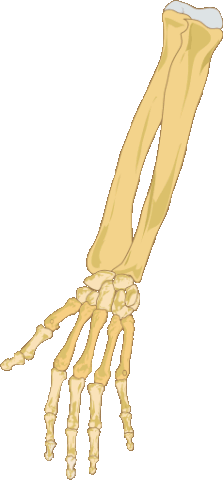
The two bones of the forearm are the radius and ulna. In the anatomical position, the radius lies on the outer side of the forearm and the ulna on the inner side.
To make it clear and memorable, the bone that aligns with our thumb is the radius bone and the bone that aligns with our little finger is the ulna bone.
They both lie parallel, and the upper end takes part in elbow joint formation, and the lower end forms the wrist joint.
Common Fractures of the Forearm: Radius and Ulna
- Monteggia fracture dislocation.
- Colle’s fracture,
- Smith fracture.
When a radius or ulna fracture near the wrist is significantly displaced, surgical fixation is recommended to restore alignment. A 2025 trial found slightly better function with surgery compared to casting in these displaced cases3.
Conclusion: Understanding Your Arm’s Structure
The powerful humerus forms the core of the upper arm and enables a wide range of motion at the shoulder. In the forearm, the pair of radius and ulna provides stability and allows for the rotation that positions our hand.
Understanding the anatomy of these bones—the humerus, radius, and ulna helps us clarify the nature of common injuries, from shoulder dislocations to Colle’s fractures. Knowing this fundamental structure is the first step toward effective treatment and a safe recovery.
FAQ

The author is a physiotherapist who has been practising for the last 17 years. He holds a Bachelor's in Physiotherapy (BPT) from SVNIRTAR (Swami Vivekananda National Institute of Rehabilitation and Research), one of the prestigious physiotherapy schools in India.
Whatever he learns dealing with his patient, he shares it with the world through blogs and e-books. He also owns a YouTube channel, "Sunit Physiotherapist" with over 8 lakh active subscribers. Here, he shares everything he gets to learn serving the patient.




Pingback: 9 Easy Supracondylar Fracture Humerus Physiotherapy Exercises| Elbow Fracture - Physiosunit
This literary analysis essay example is a nice help for everyone interested in writing craft and wish to find answer on all the interesting related issues.
That first diagram picture of a humerus is incorrectly labeled as a femur. "Greater Trochanter" should be "Greater Tubercle". "Medial and lateral malleolus" should be "medial and lateral epicondyle". There is no such thing as a "condylar". That line is pointing to a random spot between the trochlea and the capitulum. Very misleading image.